Bone Marrow and MHC/HLA Molecules

There are two main types of bone marrow transplants discussed in the literature, autologous and allogeneic. Autologous transplants are performed by using the patient’s own stem cells, collected from the bone marrow, and then reinjected back into the patient. This is done because chemotherapy/radiation therapy can kill bone marrow and blood-producing cells. Allogenic transplants, on the other hand, use cells from a donor rather than preserving and administering the patient’s own cells after a procedure. One benefit of using this method is the graft-versus-tumor effect, which is when the donor’s T cells attack the patient’s tumor.

As mentioned previously, the first part of this process is where stem cells are collected from the blood or bone marrow, depending on the patient’s current health status. To prep the patient for this procedure, the patient then undergoes chemotherapy/ radiation therapy. This preparation will not only damage/ destroy the disease, but it also acts to pacify the immune system so that the later transplant is not rejected by the body. Next, cells, whether they are from a donor or the patient themselves, are injected into the bloodstream. From here, cells travel to the bone marrow, where they attach to other cells and begin to reproduce the cells lost during the chemotherapy treatment.

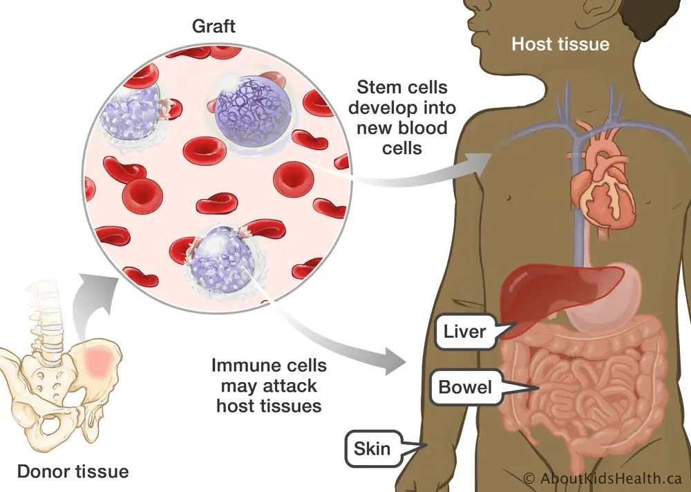
Image created by AboutKidsHealth.ca

A match is considered to be 10/10 if the donor and recipient have identical alleles present at all 5 HLA (human leukocyte antigen) loci. If the donor and recipient are not 10/10 matches, then the chance of complications rises; one example of this is graft-versus-host-disease (GVHD), where the grafted cells attack the patient’s body. You are most likely to find a 10/10 match by looking to a patient’s siblings (specifically full siblings). If this is not possible, then the most valuable resource for finding a match is the donor registry, where many different doses are preserved in case a future patient can not find a match through normal methods. Because of this, it is important for as many people as possible to donate, as it increases the likelihood of a match being found for patients with no families.

A haploidentical match occurs when a donor only matches half of the patient’s HLA alleles. This means that these types of matches only share 1 haplotype, which is where the name originates. This match is less preferable than the 10/10 match, but the advantage is that it is much easier to find this match type among the patient’s family as well as in the donor registry. This match is always found in the form of parents and children of the patient, but also with siblings at a lower rate. In the past, haploidentical matches showed a much higher complication rate, but with the use of modern technology, the chances of these events occurring are much lower.

GVHD is a common complication for patients undergoing a bone marrow transplant procedure. It occurs when a stem cell transplant attacks the host’s tissues rather than taking root properly. Specifically, it is the donor’s T lymphocytes that become activated when they contact the host cells’ antigens presented by HLA molecules. This can cause complications if the patient is not a 10/10 match with the donor, as the donor cells may recognize these molecules as invasive pathogens and attack them. This can occur in both types of bone marrow transplants, but it is more commonly seen in allogeneic transplants. In cases of severe GVHD, patients may experience both acute and chronic symptoms. Acute symptoms include rash, jaundice, vomiting, and diarrhea. Some chronic symptoms are scarring around the affected area, dry glands, muscle aches, organ damage, and possibly death.

The more HLA mismatches that are present between a donor and patient, the more likely the chances of negative outcomes. This is because it provides the donors’ T lymphocytes more chances to encounter mismatched HLA cells and attack them as foreign pathogens. Several treatments have been developed for GVHD; these include medications like corticosteroids and general monitoring and treatment of symptoms as they arise. The most effective treatment for treating GVHD, however, is still prevention. This is done by suppressing the immune response with various medications while still in the conditioning phase of a bone marrow transplant.

Inflammation and the Microbiome Part 2

Studies have shown that high-fat diets can disturb the natural gut microbiota in a way that allows for the establishment and growth of harmful bacteria, like Bilophila wadsworthia, which occupy the vacant niches left in the gut by a poor diet. As these microbes form colonies, they activate the receptors to produce lipopolysaccharide (LPS), which causes prolonged activation of the immune system. Over time, this deteriorates the gut barrier, which allows bacteria and their products to enter the bloodstream. Once that happens, the body activates a prolonged immune response to fight the constant influx of bacterial matter. If the source of invasion is not remedied or the bacteria are not otherwise eliminated, the immune system will exist in a chronically inflamed state.

As shown in the study by Sokol et al., much of the resulting inflammation is driven by activation of the TLR4 pathway, which is a direct response to the accumulation of LPS. The TLR4 pathway is responsible for later initiating downstream cytokine signaling. When TLR4 is activated by the presence of a pathogen, it increases the production of cytokines TNF-α, IL-6, and IFN-γ. These cytokines will work to remove harmful bacteria from the gut, as well as having an impact on other systemic processes, such as the regulation of blood-glucose levels. In the study presented, mice were exposed to both the high-fat diet and a normal diet, and the mice on a high-fat diet showed increased levels of B. wadsworthia, impaired glucose tolerance, and increased liver fat. This study also reported that using a probiotic to reduce the number of Bilophila wasdworthia led to decreased systemic stress from signaling.

As previously mentioned, a high‑sugar diet can reshape the gut microbiota in ways that cause inflammation. In this experiment, mice that were fed a high sugar diet developed more severe colon inflammation than those on a non-supplemented diet. In particular, the sugar diet promoted the growth of bacteria responsible for the destruction of mucus-producing cells. Some of the common species responsible are Akkermansia muciniphila and Bacteroides fragilis. As mentioned previously, this can lead to exaggerated immun activation due to the epithelium of the gut being exposed to an increased number of pathogens without the protection of mucus. These types of organisms must be eliminated or at least lowered in number so that they cannot produce LPS that damages the gut lining

This study also showed that a high‑sugar diet reduced the population of butyrate-producing bacteria, which are important for maintaining the gut barrier. The resulting decrease in butyrate means increased inflammation of the gut epithelium and the immune system, which weakens the regulation of TLR and NLR signaling pathways. As previously mentioned, lower butyrate levels cause epithelial and myeloid cells to become more exposed and sensitive to particles like LPS. This leads to heightened TLR4 pathway activation, which in turn causes downstream NF-kB signaling. This prepares the NLRP3 inflammasome for release by macrophages or dendritic cells, promoting IL‑1B production, which leads to inflammation. This study does a good job of showing how a high-sugar diet can cause multiple problems that exacerbate each other unless the source of dysbiosis is addressed.

Dietary fiber, as described in this study, is used in the process of fermentation by gut microbiota to produce short-chain fatty acids (SCFAs). These cell products are crucial for controlling immune processes. SCFAs act through multiple mechanisms to alter the gene products of cells in the immune system, nervous system, and intestines. Butyrate and other SCFAs promote the production of regulatory T cells (Tregs) and increase the secretion of anti-inflammatory cytokine IL‑10. SCFAs also support antigen-presenting cells and macrophages, which in turn reduces their output of pro‑inflammatory cytokines like IL-6 and TNF‑a.

As suggested by the studies above, increasing dietary fiber can help correct dysbiosis by promoting the growth of normal bacteria in the gut. This will lead to the establishment of SCFA-producing bacteria. The return of these organisms to normal levels and the influx of available SCFAs will allow for repair of the intestinal barrier and decreased inflammation. A high-fiber diet supports Treg production and activity, as well as anti-inflammatory cytokine production. The studies above all imply that shifting from a low-fiber, high-fat/sugar to a high-fiber, low-fat/sugar diet can reverse microbial imbalances and help to support the recovery of damaged cells.

The Role of Complement in Lupus

As described in this overview, the classical pathway has four primary functions, and each of them are activated by different molecules. The pathway is triggered when the C1 complex binds to the Fc section of an antibody that is attached to an antigen on the surface of a pathogen. This activation leads to the cleavage of complement proteins, eventually forming C3 convertase, which is a complex containing C4 and C2. The C3 convertase then produces C3a and C3b. C3b is responsible for binding to the surface of a pathogen to allow for opsonization to occur with the aid of phagocytes

C3a can also be used in combination with C5a to create anaphylatoxins. These cause inflammation and activate mast cells, as well as attracting neutrophils (also known as PMNs) to the infection site. The complex of C5b and proteins C6, C7, C8, and C9 assemble to create the membrane-attack complex (MAC). This complex causes lysis of many different types of microbes and is most effective against extracellular bacteria. The classical pathway is thus especially effective against extracellular pathogens, such as bacteria in the bloodstream that have been marked with antibodies for destruction..

Systemic Lupus Erythematosus (SLE) is a chronic autoimmune disease in which the immune system produces antibodies against itself, causing inflammation and tissue injury. Abnormalities in the classical complement pathway cause both the development and progression of SLE. Individuals who are deficient in early complement components, like C1q or C4, are more likely to develop Lupus due to impaired elimination of apoptotic cells and immune complexes as they form. This error allows the self-attacking antigens to proliferate and stimulate the production of autoantibodies. These complement deficiencies diminish tolerance, which causes the autoimmune inflammation characteristic of lupus.

During active lupus flares, the same complement system that normally protects the body creates inflammation and damages the organs. Overactivation leads to the formation of abnormal complement products such as iC3b, C3dg, and C4d, which mark immune activation and can be accurately scaled with disease severity. These products promote vascular inflammation, thrombosis, and can damage organs such as the kidneys, joints, and central nervous system. The MAC also forms on host tissues, causing tissue damage through lysis and prolonged inflammation. Through these pathways, complement proteins can cause both the start of SLE by an error in normal production, and their overactivation fuels ongoing cell damage throughout the nervous system and in various organs.

The classical complement proteins found at low levels in Lupus cases are C1q, C4, and C2. These low levels can occur either because of inherited genetic deficiencies or because the proteins are destroyed during ongoing classical pathway activation by overstimulated immune complexes. As described in this article, measuring these proteins helps to assess disease case severity and anticipate potential lupus flare-ups. The MAC contributes to organ injury by directly causing cell lysis. Immune complex deposition through C3b and C4b damages tissues such as the kidneys, joints, and nervous system, as discussed earlier. There are also the anaphylatoxins C3a and C5a (see above), which recruit immune cells and increase vascular permeability, prolonging the disease.

Luckily, new therapies are being developed to target complement activation in lupus. One developing technology uses C5 inhibitors to block the MAC and reduce tissue damage caused during flares of inflammation. Another technique aims to block C3 or parts of the classical pathway to limit inflammation. These treatments try to reduce the harmful effects of complement while keeping its protective functions intact, as that is also a contributing factor for the onset of Lupus. Early studies suggest that these therapies could help manage the symptoms of Lupus in the future.

mRNA Vaccines

Katalin Kariko and Drew Weissman are the recipients of the 2023 Nobel Prize in Physiology or Medicine. While mRNA medicines had been researched for years, they could not be used at the time because synthetic mRNA often caused dangerous immune reactions from the body, similar to the reaction catalyzed by contact with a virus. By replacing the RNA building block “uridine” with a modified version called “pseudouridine,” they avoided triggering an immune response. This change also made mRNA more stable and improved its ability to produce proteins inside cells. Their work solved core issues that had kept mRNA technology from being implemented in healthcare.

Because of their discoveries, vaccine development changed rapidly. Kariko and Weissman’s methods allowed for the streamlined design of vaccines that only required the genome of the virus they are targeting. Notably, this technique was then used to create the Pfizer and Moderna COVID-19 vaccines. Even before the pandemic, their research had already inspired new directions in treating diseases through vaccines and protein therapy. These breakthroughs have made the overall process of vaccine research and development faster and more effective overall.

MERS-CoV and SARS-CoV-2 are members of the betacoronavirus family that both use the (S) spike protein to enter host cells. If these spike proteins can somehow be damaged or rendered useless, then the viral particles will be unable to create an infection. For these reasons, researchers were aiming to create a vaccine to neutralize the viral spikes. According to this article, prior research on MERS-CoV allowed scientists to understand how to stabilize the spike protein in its prefusion shape, which is what triggers an immune response. There is a “2P” mutation that naturally presents itself in MERS-CoV that stabilizes the spike protein. This mutation was transplanted onto the SARS-CoV-2 spike, rendering the same effect as in its original organism. The researchers were then able to adapt the mRNA platform for COVID-19 once the new viral sequence was isolated.

Vaccine trials also guided the safety and dosing strategies used for COVID-19 vaccines. The article mentioned above reported that vaccine experiments using lipid nanoparticles (LNPs) demonstrated that mRNA vaccines could provide strong immune protection without severe side effects. These findings gave researchers confidence that mRNA was a safe and effective platform for coronavirus vaccines. The same study also shows how “prototype pathogen” preparedness allowed researchers to move through phases of experimentation and clinical trials at an unheard-of pace. This article suggests that the combination of prior MERS research, validated vaccine designs, and pandemic planning directly enabled the rapid and successful creation of COVID-19 mRNA vaccines.

Israeli research consisting of a large, nationwide health-care database to compare vaccinated individuals with matched unvaccinated controls. Their study found that vaccinated people had significantly lower risks of infection, hospitalization, and COVID-19–related death than those who did not get vaccinated. Because COVID-19 was a leading cause of excess mortality at the time, it was concluded that vaccination reduced both COVID-specific and overall mortality in the population studied. Reports indicated a small increased risk of myocarditis after vaccination, estimated to be somewhere between 1 and 5 cases per 100,000 vaccinees in the short period following vaccination. This data validates the claim that myocarditis is one of the adverse events associated with the vaccine. The study also notes that for males between the ages of 16 and 30, the prevalence may be closer to about 10 cases per 100,000.

These researchers compared the small excess risk of myocarditis after vaccination with the much higher risks of serious outcomes, including myocarditis, following SARS-CoV-2 infection. They concluded that the overall safety profile of the “BNT162b2” vaccine was favorable and that its benefits in preventing infection, hospitalization, and COVID-19 death outweighed the relatively rare risk of myocarditis. Most post-vaccine myocarditis cases were mild and resolved with standard care. The study emphasizes the importance of continued monitoring and clinical management of myocarditis as a rare adverse event. Overall, the authors support vaccination while acknowledging the need for vigilance regarding uncommon cardiac complications.

Misuse of Antibiotic Therapy on Children

Early antibiotic use in childhood appears to carry a measurable risk for later respiratory and allergic outcomes. In this study, which included 1,401 US children, antibiotic exposure during the first six months of life was highly associated with a 1.5 times greater chance of asthma by age six, as well as having similarly increased odds for developing allergies. The authors specifically found that this association persisted even among children without early lower‐respiratory tract infections and among those whose asthma onset occurred after age three, which reduces the likelihood that the association is simply reverse causation. An example of this error would be the early onset of asthma (wheezing) being treated with antibiotics after the fact.

The study centers on this early window (first six months) and suggests that antibiotic exposure during this period of immune system development may disrupt the normal development of the microbiome, impairing the development of immune tolerance and thereby increasing the risk of allergies or asthma later in life. Microbiome studies indicate that broader-spectrum agents may have greater disruptive potential. These findings underscore that when antibiotics are considered for use in very young infants, especially multiple courses or broad-spectrum agents, health care professionals and parents should weigh the risk of long-term immune system function risk alongside the immediate need for treatment. Further studies have suggested that broad-spectrum antibiotics, particularly macrolides and cephalosporins, are most strongly associated with increased asthma and allergy risk. These classes are thought to have greater disruptive effects on gut microbial diversity, which may explain their stronger link to immune-related outcomes.

Early antibiotic exposure is also linked to higher risks of several childhood gastrointestinal disorders, such as inflammatory bowel disease (IBD), and some studies indicate celiac diseases and functional disorders such as IBS as well. This review found that exposures in the first years of life were associated with later IBD diagnoses, with some studies indicating that more courses of antibiotics increase the risk proportionally. Associations for celiac disease and IBS tended to follow the same pattern, especially when antibiotics were given early in life. The authors emphasize that the strongest signal appears when antibiotics are given during the important microbiome development period discussed earlier. The review concludes that early-life antibiotic use may increase later GI disease risk and that, as stated before, medical professionals and parents should exercise caution when giving their young children antibiotic treatments.

Regarding antibiotic types and mechanisms, the review highlights that broad-spectrum agents (those that substantially disturb microbial diversity) are most often implicated in the appearance of these conditions, whereas more narrow-spectrum agents (such as penicillin) do not show a consistent link. The proposed mechanism is microbiome disruption during the previously mentioned critical developmental window, leading to long-term changes in immune regulation and gut barrier function that can predispose to immune-mediated gut disease. It is important to note the limitations of the source studies, as they are mostly observational and may not account for other factors, such as “confounding by indication”. This means that the infections that prompt the use of antibiotics in infants and young children may also themselves increase the chance of disease later in life. In summary, early and repeated broad-spectrum antibiotic exposure is associated with increased risk of later GI disorders, but more studies would need to be conducted to rule out other possibilities

In the United States, a significant proportion of outpatient antibiotic prescriptions for children are unnecessary, particularly for conditions resulting from viral infections. It is estimated that about 90% of otitis media/bronchitis cases are caused by viral infections. The Centers for Disease Control and Prevention (CDC) also estimates that at least 60% of the time, antibiotics are prescribed for these infections. To reduce inappropriate antibiotic prescriptions for viral infections in children, the CDC has implemented outpatient programs that provide guidance on when it is wise to prescribe antibiotics to children. These programs include education, support tools, and feedback systems that track prescribing patterns and encourage adherence to CDC guidelines. Public awareness campaigns also aim to educate parents and caregivers about when antibiotics are necessary, which may act as a second barrier between providers who are financially incentivized to prescribe drugs and the children who take them.

The research described above does indicate that inappropriate antibiotic prescriptions for viral infections in early childhood likely contribute to the problems described above. All of this indicates that unnecessary antibiotic use can disrupt the developing gut and airway microbiomes during crucial periods of immune system development, which is linked to increased risks of asthma, allergies, and GI disorders like IBD or IBS. Broad-spectrum antibiotics seem to cause the greatest disruption to normal development by reducing overall microbial diversity, which may change immune regulation long-term. While observational studies cannot stand on their own to provide a definitive link, the consistency of associations across multiple studies suggests that unnecessary and/or prolonged antibiotic exposure is a significant risk factor. Reducing these prescriptions should be done, not only because it limits immediate side effects and resistance, but may also help prevent chronic immune-mediated conditions that arise later in childhood or adult life.

Antibiotic Resistance and the NDM-1 Plasmid

The NDM-1 plasmid encodes a bacterial enzyme called the “New Delhi metallo-β-lactamase” enzyme, which gives certain bacteria the ability to break down most β-lactam antibiotics. The NDM-1 plasmid also carries other genes responsible for antibiotic resistance. The NDM-1 gene was first found in plasmids of Klebsiella pneumoniae in India. The gene has since spread worldwide and is now found in many habitats, with a typical reservoir for infection being hospitals. Through the process of conjugation, in which bacteria use a pili to transmit a gene, resistance can be transmitted to new bacteria. For these reasons, infections caused by NDM-1–producing bacteria are particularly difficult to treat and represent a growing public health concern as their resistance continues to adapt to existing treatments.

Recent surveys in the United States have revealed a sharp rise in NDM-1–producing CRE (carbapenem-resistant Enterobacterales) infections. This research shows that in New York City, reported cases increased from 58 in 2019 to 388 in 2024. This indicates increased rates of both local transmission and global introduction of resistant strains from abroad into public spaces, especially hospitals, as mentioned before. A CDC study found that NDM-CRE infections rose by about 460% between 2019 and 2023. These results showcase the need for better infection control and surveillance to mitigate the spread of NDM-1 resistance in the U.S.

Image created by Li et al.

One of the main reasons that pharmaceutical companies often opt for producing drugs to treat chronic illnesses rather than antibiotics is that it is more cost-efficient. The results of this study show that antibiotics are more often prescribed for short-term durations and are usually only used as a last resort to prevent resistance from building in bacteria over time. This ultimately limits profit potential and is negative for the pharmaceutical companies. This is because long-term treatments usually cost more than short-term ones and result in more profit for shareholders. These incentives, among others, received by hospital staff, all contribute to the overuse of antibiotic treatments.

Developing new antibiotics is challenging beyond financial pressures. The process requires extensive, time-consuming testing to ensure the drugs are safe and effective, which takes considerable time and resources. At the same time, hospitals and doctors carefully limit the use of new antibiotics to prevent resistance, which reduces overall demand. While these stewardship practices are important for public health, they make it more difficult for companies to justify the investment. While doctors and other healthcare workers do their part to stop resistance from building, there is only so much that can be done in the face of such financial pressures.

Image provided by Centers for Disease Control 2011 Data

A study published in the New England Journal of Medicine investigated how often viruses contribute to acute otitis media in children. In this study of 456 pediatric patients, researchers found that over 40% had viral pathogens present in their middle-ear fluid. This finding shows that a large portion of ear infections traditionally treated with antibiotics may actually be viral. Because antibiotics do not treat viruses, this suggests that many prescriptions in these cases may be unnecessary and could lead to increased treatment resistance in new strains of bacteria. These results highlight the importance of careful clinical evaluation before beginning antibiotic therapy, and more temperate use of antibiotics in general.

The same study emphasizes that viruses such as RSV can directly infect the middle ear, further supporting the idea that many infections resolve without antibiotics. These findings imply that medical professionals may overuse antibiotics when viral causes are not considered, possibly due to financial incentives, as they are criticized for broadly prescribing antibiotics. This raises broader concerns about how unnecessary antibiotic exposure may disrupt the developing microbiome. A 2021 systematic review reported that early-life antibiotics are associated with a higher risk of childhood IBD and celiac disease. The conclusion of this review was that these associations are likely linked to microbiome disruption during critical developmental windows for natural, healthy bacterial growth. This suggests that unnecessary antibiotic use for viral infections may contribute to later gut complications, as well as allergies and mental disorders.

MMR Vaccines and the Body

Andrew Wakefield published a paper in The Lancet in 1998 that included 12 children claiming that the MMR vaccine was positively linked to autism. The children were all patients of the Royal Free Hospital in London, being treated for gastrointestinal and developmental issues. Data was collected utilizing the parents of the patients’ memories, and there was no control group in the study. Flaws like these, as well as ethical violations, forced the papers’ retraction as well as Wakefield’s medical license being revoked. The publication of this paper caused an uprise in vaccine hesitancy, and vaccination rates dropped, leading to outbreaks of diseases that had previously been under control.

After this study was published, many independent research groups investigated the supposed link between the MMR vaccine and autism but found no supporting evidence. The Institute of Medicine reviewed numerous large-scale epidemiological studies and concluded that there is no causal relationship between the MMR vaccine and autism. Their report emphasized that autism symptoms typically appear around the same age vaccines are administered, which may have led some parents to mistakenly associate the two. This conclusion has been reinforced by additional studies worldwide, all showing no increased autism risk among vaccinated children. Today, Wakefield’s study is widely recognized as fraudulent, and the overwhelming scientific consensus confirms that vaccines do not cause autism.

Image credited to CNN

In Denmark, the national vaccination program administered the MMR vaccine to all children at around 15 months of age. In 2002, Madsen et al analyzed data from over 537,000 children born from the years 1991-1998 to examine the relationship between MMR vaccination and autism. Children who had not received the vaccine served as the control group, and the researchers controlled for variables such as gender, birth weight, and socioeconomic background. By using comprehensive registry data, the study avoided issues like recall bias and selective sampling, which was one point of criticism of the Wakefield paper. The purpose of the research was to determine whether there was a link between receiving the MMR vaccination and the onset of autism.

Wakefield’s 1998 study, unlike Madsen et al’s study, examined only 12 children who had developmental regression and gastrointestinal issues and were all treated at the same hospital discussed earlier. Also, unlike the study above, this one relied on parents’ abilities to remember when symptoms of autism began. The study’s claim was that symptoms often appeared shortly after MMR vaccination, but the small sample and lack of control or comparison group made it impossible to establish cause and effect between autism and the MMR vaccine. The study was also limited by selection bias, as the children were not randomly chosen and were all from the same location and may share other qualities; therefore, they may not be representative of the greater population. In contrast to Madsen et al.’s large, nationwide analysis, Wakefield’s findings were unreliable and ultimately discredited by further research showing no link between the MMR vaccine and autism.

Image Created by AutismBC

Relative risk (RR) is a statistical tool used in public health and epidemiology to determine how likely an event is to occur in one group compared to another. It is calculated by dividing the rate of the outcome in the exposed group by the rate in the unexposed group. An RR of 1 indicates no difference in risk between the two groups, an RR above 1 means the exposure increases the risk, and an RR below 1 suggests a protective effect. This measurement is particularly helpful in vaccine studies or other health research to quantify whether an exposure changes the probability of a specific outcome. An understanding of RR is crucial for many roles in public health, academia, and policy-making.

The Madsen et al study used relative risk to evaluate whether the MMR vaccine contributed to autism. They reviewed health records for over half a million children born in Denmark from the same period of time as the original study, comparing autism rates between those who received the vaccine and those who did not. The study reported an RR of 0.92 with a 95% confidence interval of 0.68 to 1.24, indicating that vaccination did not increase the chance of developing autism. This shows that there were similar rates of autism among all children, whether they were vaccinated or not, refuting all claims of a cause-and-effect relationship. Overall, this retrospective study not only refutes the claims of Wakefield but also serves as proof of the safety of MMR vaccines.

Ultraprocessed Foods and the Body

Ultraprocessed foods are food products that have been altered, usually in order to extend shelf life and/or make them more palatable. This is accomplished through multiple rounds of processing, as well as the use of additives such as artificial colors, flavors, and preservatives, among other substances. Some products that fit into this category are soft drinks, snack foods, frozen food, chips, etc. These foods often contain extra ingredients such as chemical additives, modified starches, and added oils, as described here. Ultraprocessed foods not only contain these added ingredients, but they also often have an overall lack of nutritional value due to a lack of vitamins, minerals, etc.

Some additives, described here, are particularly common in a wide variety of foods. Emulsifiers are added to foods like ice cream, chocolate, and mayonnaise to keep ingredients mixed and to give them a smooth texture. Artificial sweeteners are often used in diet sodas, yogurts, or protein powders to provide sweetness while not including sugar. Preservatives are included in things such as packaged breads, canned goods, or processed meats to slow spoilage and extend shelf life. To make products more appealing, colorants like Red 40 give foods a consistent look, while flavor enhancers like MSG intensify taste in snacks, soups, and instant noodles. Together, these additives help processed foods stay stable, look attractive, and taste the same every time you buy them.

Photo by Evan-Amos

Short-chain fatty acids (SCFAs) are important byproducts of gut microbial fermentation, and recent human research shows that emulsifiers can lower their production. In a double-blind randomized trial, healthy adults first consumed an emulsifier-free diet and then were given brownies containing one of several emulsifiers, such as carboxymethyl cellulose, soy lecithin, etc. Across the groups, SCFA levels dropped compared to placebo, most strongly in the carboxymethyl cellulose group, suggesting a negative impact on microbial metabolism. Overall, microbial diversity did not change much, but their function was impaired. This finding highlights that emulsifiers can alter microbes’ ability to function in their common role, even when the number of species remains stable.

The same study also looked at how these additives shift gut microbial composition. While it didn’t report major across-the-board decreases in Bifidobacterium or Faecalibacterium, earlier studies have shown that these beneficial taxa can be reduced by emulsifier exposure. Because both genera are known for producing butyrate and supporting anti-inflammatory processes in the gut, even small changes in their activity could explain the lower SCFA levels found in participants. The trial’s results suggest emulsifiers may quietly weaken beneficial microbial functions without causing dramatic changes in overall diversity. All of this data goes to show how ultra-processed food additives can negatively impact gut health.

Image by Lee et al

Research suggests that consuming ultra-processed foods (UPFs) may increase both the risk and severity of multiple sclerosis (MS). A study found that each extra daily serving of UPFs was linked to a higher chance of central nervous system demyelination, and higher overall intake was associated with nearly triple the odds of moderate-to-high MS severity. UPFs typically contain refined sugars, saturated fats, trans fats, and high levels of salt, which can promote inflammation and disrupt the balance of gut bacteria. This imbalance may reduce the production of SCFAs and increase intestinal permeability, contributing to systemic inflammation and immune system dysregulation. Overall, these factors suggest that diet plays a significant role in MS development and progression.

Focusing on whole-food diets may help mitigate the negative effects of UPFs on MS. Diets rich in fruits, vegetables, whole grains, and healthy fats support gut health and reduce inflammation. Maintaining a diverse and balanced gut microbiota helps preserve SCFA production, which is important for regulating immune responses. Limiting UPFs can also reduce activation of pro-inflammatory T helper cells, which are implicated in the onset of MS. In summary, replacing ultra-processed foods with nutrient-dense whole foods could be a practical approach to managing MS risk and symptoms.

Methylmercury and the Body

Methylmercury is an organic compound that is generated by some aquatic microorganisms. This occurs so that the organisms may convert naturally occurring mercury into a form that is useful for necessary biological processes. This new compound is stored in body tissues, where it binds with muscle proteins of aquatic animals. While this process may occur in microorganisms, these will be eaten by larger organisms that store methylmercury in their tissues as well. Eventually, this could lead to humans ingesting these animals and absorbing the methylmercury as well.

One common source of Methylmercury in humans is tuna. Different subspecies of tuna contain different amounts of this compound due to their behaviors and diets, as discussed in this paper. Light tuna, for example, are smaller fish with shorter lifespans, so they feed lower on the food chain and ingest less methylmercury. Albacore tunas are slightly larger and live a more predatory lifestyle than the light tuna, so they have higher methylmercury levels. Sushi-grade tuna are the largest, longest-lived, and most predatory among this group and therefore have the highest levels accumulated from consuming other organisms.

Photo of Tuna by NOAA Fisheries

As stated here, the signs and symptoms of methylmercury build-up are numbness of the fingers or limbs, impaired coordination and/or strength, vision/hearing impairment, slurred or otherwise altered speech, as well as cognitive decline. Severe and/or prolonged cases can lead to permanent developmental delays in children, as well as seizures or even death. These symptoms typically develop gradually over time. This is because Methylmercury is classified as a neurotoxin, which is specifically designed to attack the nervous system. Young and unborn children are therefore more at risk for injury since their underdeveloped nervous systems are more vulnerable to these toxins.

Because of the risk of Methylmercury in large amounts, the FDA has issued guidelines for tuna consumption. For the normal, healthy adult, the FDA recommends and serving size of about 4 ounces. However, exceptions to this recommendation exist due to increased risk for vulnerable groups. For pregnant women, only about 2 or 3 servings a week are recommended, and only 1 if they are eating albacore tuna. Pregnant women should totally avoid sushi-grade tuna. For children, the recommendation varies by age, with the youngest children (ages 1-3) only recommended to eat a 1-ounce serving twice a week. On the other end of the spectrum, college-age adults can consume up to 3 full-sized (4-ounce) servings a week.

Albacore Tuna photographed by NOAA Fisheries

Thimerosal is a compound used as a preservative, popularly used in vaccines in order to prevent bacteria or fungal organisms from contaminating the compounds the vaccine is attempting to deliver. Once inside the body, it is broken down into ethylmercury and thiosalicylate. Both are circulated through the blood, and ethylmercury (which remains the longest) is eliminated from the body in a maximum of 10 days. It is an organomercury compound, and is about 50% mercury. This relates the compound back to our previous topic of methylmercury, which has been cited for its neurodegenerative effects. Some have speculated that thimerosal may be harming patients who are treated with vaccines containing this preservative.

One 2011 study found that cases of atypical autism diagnoses were “statistically significantly more likely” to have higher cumulative thimerosal exposure, which seems to support this theory. However, another article from 2011 found that epidemiologic evidence does not support the relationship between vaccines containing thimerosal and autism diagnoses. This article reviewed epidemiologic studies published between 2003 and 2008 and found that while some did show a positive relationship between the two factors, the relationship wasn’t strong enough, and it did not occur in enough studies to be a reliable trend. Overall, the consensus outside of these studies is that thimerosal is not linked to autism. While some studies over the years have found a link, there have been criticisms over their methodology.

Finally, my favorite kind of tuna is albacore. I like to eat them in a toasted sandwich with cheese. I can’t imagine that I eat more than 4 ounces a week, so I believe I am safe from methylmercury specifically. The studies indicate that you should be more cautious of mercury from tuna than vaccines, as there are more confirmed incidences with tuna, and the vaccine risk is not confirmed.

The Microbiome

The microbiome exists in all humans, and it plays a crucial role in many processes throughout the body. The main role of the microbiome is to be the regulator of the gut-brain axis, as is described in this article that investigates therapeutic applications for the axis. The microbiome also serves in other roles, such as producing some valuable substances as byproducts of its activities. One example of this is how gut microbes produce short-chain fatty acids, which are important for neurological processes. The immune response and strength are also heavily influenced by the microbiome.

The Gut-Brain axis is a bidirectional network of communication that runs between the brain and the microbes living within the gut. This axis allows the nervous system to respond to changes detected in the gut microbiome as well as vice versa. This ensures that both systems are successfully maintaining homeostasis and are able to make changes to support the other. One example that illustrates how important the relationship between these two ends of the axis is to each other is the phenomenon of dysbiosis. Dysbiosis occurs when there is an imbalance of some kind with the population of microbes within the gut. This gut issue can lead to issues like neuroinflammation and neurodegeneration, which affect the brain directly if left untreated.

Image by Nakatsu et al.

As described in the article given to us, a neuropod is a type of epithelial cell that is found in the small intestine as well as a few other areas. Its function is to detect nutrients nearby and communicate this information to the vagus nerve in order to influence an animal’s appetite, thereby reducing the amount of gut nutrients in surplus or raising the amount present if it is lacking. Neuropods are also used in other parts of the body to gather information about the local environment. For instance, they are found in the colon, where their job is to detect signs of microbial molecules that could lead to an illness. Neuropods are part of a complete system that we are still learning about, which is why researchers at Duke were able to discover a new feedback system utilizing neuropods this past summer!

In the colon, where neuropods are used to detect microbial proteins, they often do so by detecting flagellin specifically. This is because flagellin is the main structural protein used by bacteria to form flagella. Neuropods are able to do this because they have receptors that are designed specifically for receiving bacterial flagella of dangerous bacteria. While there may be other microbial proteins present, neuropods have unique signal pathways for each type of protein detected. If the neuropod were to receive some microbial protein other than flagellin, its corresponding signal and effect would be completely different than the one described in the article.

Helicobacter pylori electron micrograph by Yutaka Tsutsumi

This article links dysbiosis to autoimmune diseases. This link is made in the article by demonstrating how local inflammation of the gut due to dysbiosis is commonly seen along with cases of autoimmune diseases. When the gut microbiome is imbalanced, it can disrupt the immune system and cause inflammation. Certain bacteria can trigger the immune system to attack the body, contributing to autoimmune diseases like type 1 diabetes or rheumatoid arthritis. Understanding the link between gut dysbiosis and autoimmunity could help develop therapies that target the microbiome to prevent or reduce autoimmune disease activity.

The disease I wanted to investigate was Crohn’s disease, and all of my information was sourced from here. Crohn’s disease is a long-term condition that causes inflammation anywhere along the gastrointestinal tract, from the mouth to the anus. Individuals with Crohn’s often live with symptoms like diarrhea, abdominal pain, fatigue, and unintended weight loss. While the exact cause isn’t fully understood, it’s thought to involve a mix of genetics, immune system issues, and environmental factors. The disease can sometimes lead to complications such as strictures, fistulas, or problems with nutrition. Treatment usually focuses on reducing inflammation with medication, and surgery may be needed in some cases to manage complications.